If you disable the "Active Content" in your browser you may not
be able to view the animations or videos supplied in this lab.
If prompted you should "Allow Blocked Content".
The models found in most schools, use compound
lenses and light to magnify objects. The lenses bend or
refract the light, which makes the object beneath them appear
closer.
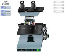
Stereoscope
This microscope allows for binocular (two
eyes) viewing of larger specimens. (The spinning microscope at
the top of this page is a stereoscope)
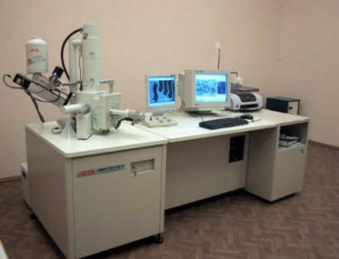
Scanning Electron Microscope (SEM)
This microscope allows scientists to view a
universe too small to be seen with a light microscope. SEMs
dont use light waves; they use electrons (negatively charged
electrical particles) to magnify objects up to two million
times.
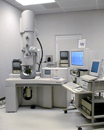
Transmission Electron Microscope
(TEM)
This microscope also uses electrons, but
instead of scanning the surface (as with SEM's) electrons are
passed through very thin specimens.
The microscope has been one of the key instruments used by the
biologist for hundreds of years. A number of improvements were
made to light microscopes during the first two or three
centuries after their invention. Since the turn of the century,
however, there have been no significant improvements. There have
been continuous advances in the methods of preparing and
analyzing specimens. There have also been modifications to the
light microscope which permit new methods of analysis (phase
contrast, fluorescence, confocal laser scanning, etc.) and the
introduction of the electron microscope which can push
microscopic analysis to the molecular level.
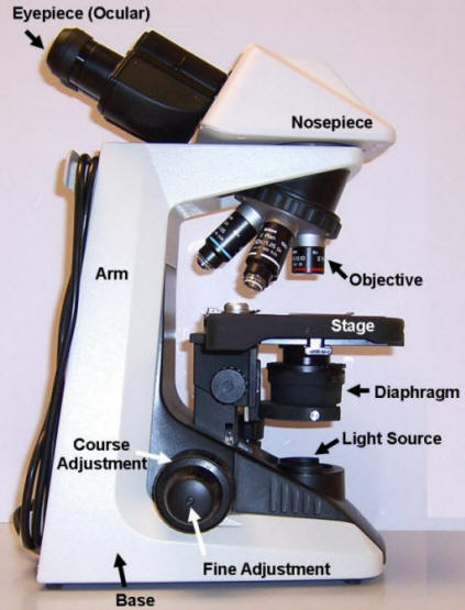
The main purpose of a microscope is to magnify and increase the
visibility of a small object. The magnification of a lens is
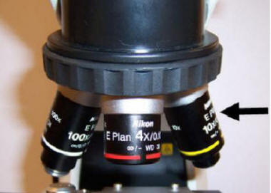
always engraved on it. You will be using a compound microscope
which has a system of lenses. The total
magnification of the scope is a product of the magnifications of
the objective lens and the eyepiece (or ocular lens). For
example, using a low-power objective (magnification = 3.4X) and
a standard eyepiece (magnification = 10X), the total
magnification is 34X.
Resolution is an important attribute of a microscope. The limit
of resolution of an optical system is the minimum distance by
which two objects can be separated and still be perceived as
distinct. Two points placed closer than this limit will be seen
as one. Greater resolution allows one to see an object more
sharply and to make out internal detail.
Adequate lighting will allow you to obtain the best resolution
possible. Illumination must be adjusted for each objective every
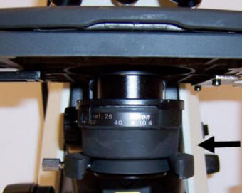
time a change is made. The adjustments which will affect
illumination on your microscope involve changing the iris
diaphragm. The iris diaphragm is used to match the aperture
(opening) to that of the objective. It should not be used to
control the intensity of illumination. With some unstained or
transparent specimens, it may be necessary to close the iris
slightly to improve contrast. This is always done at the expense
of resolution.
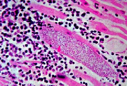
Figure 1.10 Mycobacterium leprae (stained dark blue)
are shown above in human tissue
Mycobacterium leprae, the bacterium
that causes leprosy was discovered in 1873 by Gerhard Henrik
Armauer Hansen (Bacteria
Genomes 2008). Also known as Hansens disease, leprosy has a
long history of associated stigma, which prevents people from
seeking medical treatment and often thwarts public health
intervention to curb further spread of the disease. For this
reason, public health interventions should include efforts to
reduce stigma via an educational awareness program and training
of local health officials.
Leprosy is a human disease caused by the
bacillus Mycobacterium leprae (Figure 1.10).
M. leprae is an acid-fast bacterium. As one of the
slowest growing bacteria known and its inability to grow
independently, successful in vitro cultivation has never been
achieved. Although found in the same genus as the tuberculosis
bacterium (Mycobacterium tuberculosis), the two
diseases cause different symptoms.
It is hypothesized that
M. leprae infects a new host by way of skin or
upper respiratory tract, but most experiments suggest the latter
as the more likely possibility. M. leprae causes a
chronic disease of the peripheral nerves, skin and mucosal
membranes of the body and has an incubation period of about 3-5
years. If initial symptoms are left untreated, then permanent
damage may result in many parts of the body including the eyes
and outer extremities.
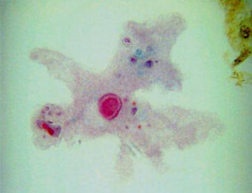
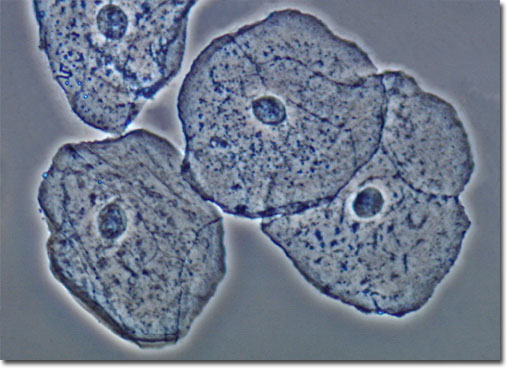
Figure 1.11
Human Cheek Cells
(The nucleus is the darker,
spherical organelle near the center of the cell)
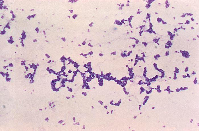
Observe the following image which illustrates the three
different types of bacteria. Use the links given below to view
microscopic slides of each of the three shapes of bacteria.
SKETCH 2 **Identify the different shapes
of bacteria in
your sketch as: Baccilus, Cocci,
Spirillum
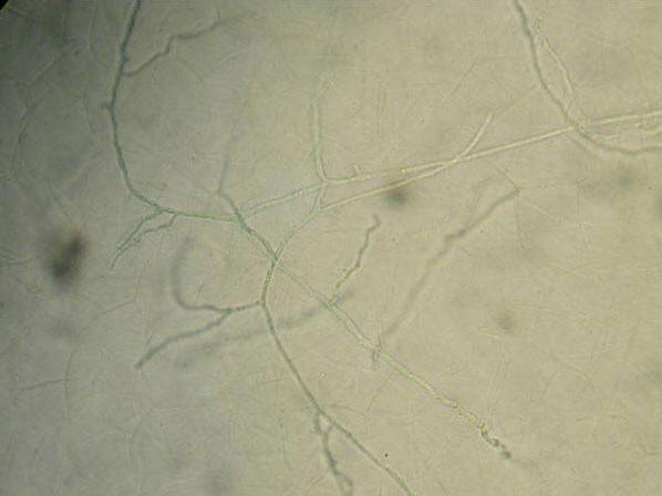
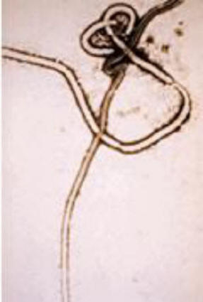
The most evident structure for most fungi is the
spore bearing mushrooms of fungi. However, the
defining character of fungi are the hyphae . Hyphae
are cylindrical, branching tubes in which the
cytoplasm of the fungus is found. Food is absorbed
through the walls from the surrounding fluids or
medium. The life cycle of fungi involve a spore (two
are shown in Figure 1.13) settling and then
the hyphae begin to grow, branching and growing
outwards. The cytoplasm tends to migrate to the
growing tips of the fungi and interconnected hyphae
may extend over many yards. Leading some to suggest
that fungi are the largest organisms on earth.
SKETCH 3 **Sketch
hyphae and spores seen in the figure to the right.
Video of a 100X microscopic
view of a drop of pond water. Click on the arrow
to view the video.
The variety of organisms you see
here would be typical of most freshwater ponds.
The small spherical organisms seen floating
about are bacteria. The large and small, fast
moving, green organisms that are darting about
are paramecium feeding on bacteria. The hat
shaped organisms are vorticella again feeding on
bacteria. The darker greenish brown masses are
bacterial colonies and algae. The green
rectangular organisms are green algae. The long,
thin strands scattered throughout the sample are
blue-green algae.
SKETCH 4 **Observe the
video and sketch some of the variety of living
organisms present
Following the mitotic process, the surfaces of the new
daughter cells seem to bubble wildly as if they were
suddenly placed under high heat and were being boiled alive.
Watch a few of the cells divide as you observe them.
1)
What is the function of the diaphragm? 2) Describe
the advantage of having a microscope of highest resolution.
3) Calculate the magnification of the lens
system of the following: a) Ocular-10X Objective-10X b) Ocular-10X Objective-43X c) Ocular-10X Objective-1X d) Ocular-10X Objective-2X 4) What is
the most important attribute of a microscope?
**Go to the following site to
link to a virtual light microscope. At the virtual
microscope site you will need to perform the
tutorial so that you learn how to use the
microscope. Click on the
GETTING STARTED link on the upper left side.
After learning how to use the
microscope and viewing the speciments, answer the questions
given below on Virtual Light Microscopy.
A) Perform the tutorial (Getting Started) so
that you learn how to use the microscope B)
Choose to view the cheek smear slide C) You
will be using the 4X,10X and 40X objective lens powers D) Use the focus and illumination slide bars to
see the cheek cells better.
E) Answer the questions on your experiences at
this site below.
You can use the page link below to access a labeled image
of the microscope
1) How many individual cells can you count at the following
objective powers of magnification?
a) 4X b) 10X
c) 40X
2) If the eyepiece has a 10X power, what is the total magnification when you observe cells
at objective power of 40X?
3) At what power are you able to discern the nucleus of the
cheek cells? (The nucleus is the large, darker organelle located near the
center of the cell)
4) Describe what happens if there is too much illumination.
Two types of
electron microscopes have been developed over the past half
century: the
Transmission
Electron
Microscope
(TEM) and the
Scanning
Electron
Microscope
(SEM).These
instruments contain magnetic lenses that focus a beam of
electrons on the specimen. Electrons used in this fashion
generate a wavelength that may be 100,000 times shorter than
that of visible light. As a result, electron microscopes have
resolving powers as much as 400 times that of light microscopes
and 200,000 times that of the human eye.
The
TEM
bombards a thin specimen with electrons. Depending on their
composition, the components of the specimen either transmit,
absorb or deflect the electrons. The image produced on a
photographic plate is a visual translation of this interaction
of electrons with the specimen. The transmission electron
microscope gave scientists their first look at the world of
viruses, invisible by light microscopy, and today permits us to
see molecules and atoms.
The
SEM
is quite different from the
TEM.
It is designed to generate three-dimensional images of surface
detail. This microscope moves an electron beam back and forth
over the surface of a metal-coated specimen causing the emission
of secondary electrons from the specimen. The secondary
electrons produce the stunning images characteristic of
scanning electron microscopy.
Video of
how an electron microscope works. Click on the arrow
to view the video.
To observe (resolve) objects smaller than 0.2 m
requires the utilization of Electron Microscopy (EM). Rather
than using visible light, electron microscopes focus a beam of
electrons on a very thin section of biological material that has
been chemically preserved (fixed) and embedded in plastic.
Electrons have a much shorter wavelength than the photons of
visible light used in LM. Since resolving power is
inversely related to wavelength, modern electron microscopes can
resolve objects of approximately 0.2 m.
It is this tremendous increase in resolution that has allowed
biologists to discern the precise details of cell structure.
Although a powerful tool, only chemically preserved cells can be
observed with EM. The routine observation of living cells
by electron microscopes is a goal yet to be achieved.
The type of electron microscopy described above is generally
referred to as Transmission Electron Microscopy (TEM). In
TEM, the beam of electrons passes directly through the
sample except where the electrons are deflected by atoms of
heavy metals (lead and/or uranium) that have been used to
"stain" the specimen; the transmitted electrons are
focused onto photographic film where the image is visualized and
recorded.
A variation on this approach is Scanning Electron Microscopy
(SEM). In SEM, the electron beam scans the surface of a
sample that has been coated with a thin layer of gold. The beam
of electrons excites the atoms of the sample causing them to
eject electrons which are collected and converted into an image
that is displayed on a monitor. The image that is produced has a
great depth of field and thus appears to be three dimensional.
SEM
is used to reveal the surface details of various types of cells.
VIRTUAL LAB
**Go to the following site to
experiment with a virtual scanning electron microscope.
Answer the questions on Virtual Electron Microscopy given
below. VIRTUAL ELECTRON MICROSCOPE
A) There are three specimens to view shown on
the left side B) You can use the MAGNIFY
button on the machine to zoom in on your specimen
C) Answer the questions on your experiences at this
site below.
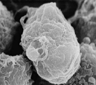
How many bacteria do you think are on
the image? 2) Describe the different
shapes of the bacteria that are visible. 3)
Which cell is the largest between the macrophage and the
bacteria? 4) Describe the shape of the
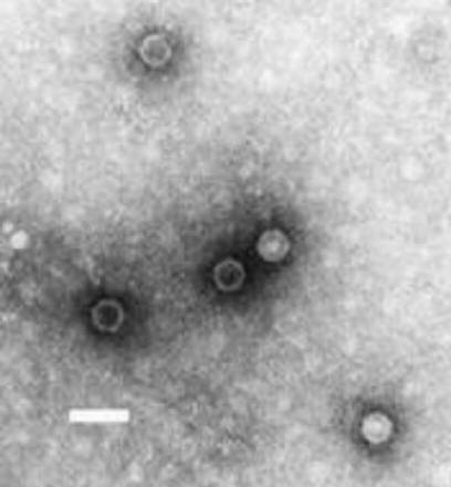
bacteria. 5) Why do you think the virus
has so many spikes on it? 6) Does the
electron microscope allow a higher degree of magnification
than the light microscope? Why?
View the Milky Way at 10
million light years from the Earth. Then move through space
towards the Earth in successive orders of magnitude until you
reach a tall oak tree just outside the buildings of the National
High Magnetic Field Laboratory in Tallahassee, Florida. After
that, begin to move from the actual size of a leaf into a
microscopic world that reveals leaf cell walls, the cell nucleus,
chromatin, DNA and finally, into the subatomic universe of
electrons and protons.